
Greetings to you . This is Emelin. Each and everytime when I'm addressing I'll be so excited because your love and support keeps me to move on further. Really thanking everyone from the bottom of my heart. keep commenting and share your ideas for my betterment .
In today's topic we are going to discuss about Tissues. But it's a vast one so I have divided it into 4 sub topics .So that it would be easier for you all . Today we are going to deal about "THE EPITHELIAL TISSUSE" . You can watch a brief explanation of these slides in both English and Tamil in my YouTube channel named "NURSING ABSTRACT".
channel link:
https://www.youtube.com/channel/UCZrabu-CtMa5AEexu1j2GNw
English video:https://youtu.be/t8TGn77BbT0
Tamil video:https://youtu.be/msN07rHpKog
INTRODUCTION TO TISSUES:
Tissues consists of a large number of same type of cells and are classified according to their size, shape , function of their constituent cells.
There are 4 main types of tissues each with subtypes .they are
1.EPITHELIAL TISSUE OR
EPITHELIUM.
2.CONNECTIVE TISSUE.
3.MUSCLE TISSUE.
4.NERVOUS TISSUE.

EPITHELIAL TISSUE & TYPES:
Epithelial tissue covers the body and lines cavities ,hollow organs and tubes.
It is also found in glands.
Its functions include protection of underlying structures from ,for example ,dehydration , chemical and mechanical damage.
Secretion and absorption.
The cells are very closely packed and the intercellular substance, matrix, is minimal.
The cells usually lie on a basement membrane , which is a connective tissue made by the epithelial cells themselves.
It is classified into
1.SIMPLE :a single layer of cells.
2.STRATIFIED: several layer of cells.

SIMPLE EPITHELIUM:
Simple epithelium consists of a single layer of identical cells.
It is usually found on absorptive or secretory surfaces, where the single layer enhances these processes.
There are mainly 3 main types of simple epithelium and are named according to the shape of the cells, which differ according to their functions.
The more active the tissue taller the cells.
The 3 main types are
1. SQUAMOUS (PAVEMENT) EPITHELIUM
2. CUBOIDAL EPITHELIUM
3. COLUMNAR EPITHELIUM.
SQUAMOUS EPITHELIUM:
This is composed of single layer of flattened cells,
The cells fit closely together like flat stones, forming a very thin and smooth membranes across which the diffusion occurs easily.
It forms the lining of the following structures
1. heart – ENDOCARDIUM.
2.blood vessels &lymph vessels- ENDOTHELIUM
3.alveoli of the lungs
4.lining the collecting ducts of nephron in the
kidney.

CUBOIDAL EPITHELIUM:
This consists of cube shaped cells fitting closely together lying on a basement membrane .
Cuboidal epithelium is involved in the process of secretion, absorption and excretion.
It forms the kidney tubules and is found in some glands such as thyroid.

COLUMNAR EPITHELIUM:
This is formed by a single layer of cells rectangular in shape ,on a basement membrane.
It lines many organs and often has adaptations that make it well suited to a specific functions.
The lining of the stomach is mainly composed by simple columnar epithelium without surface structures.
The free surface of the columnar epithelium is lining the small intestine and it is mainly covered with the microvilli . the microvilli provides a very large surface area for absorption of nutrients from the small intestine.


In trachea columnar epithelium is ciliated and also contain goblet cells that secrete mucus .this means the inhaled particles stick to the mucus layer are moved towards the throat by cilia in the respiratory tract.

In uterine tubes the ova are propelled along by ciliary action towards the uterus.
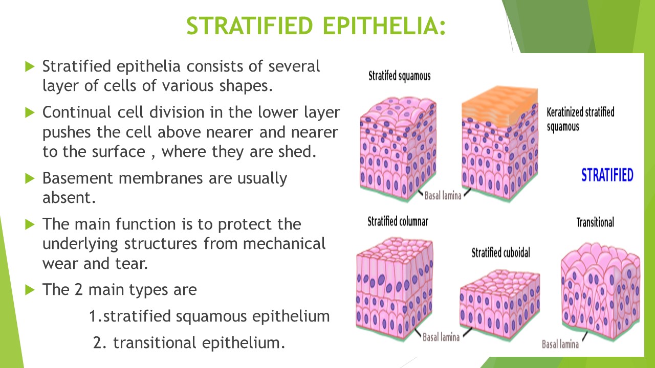
STRATIFIED EPITHELIA:
Stratified epithelia consists of several layer of cells of various shapes.
Continual cell division in the lower layer pushes the cell above nearer and nearer to the surface , where they are shed.
Basement membranes are usually absent.
The main function is to protect the underlying structures from mechanical wear and tear.
The 2 main types are
1.stratified squamous epithelium
2. transitional epithelium
.

STRATIFIED SQUAMOUS EPITHELIUM:
This is composed of several layer of cells.
In the deepest layer of the cells are mainly columnar as they grow towards the surface they become flattened and they shed.
KERATINISED STRATIFIED EPITHELIUM:
This is found on dry surfaces subjected to wear and tear i.e. skin ,hair , nails. The surface layer consists of dead epithelial cells that have lost their nuclei and contain the protein KERATIN. this forms a tough and relatively a water proof protective layer that prevents the drying of the cell underneath .the surface layer of the skin is rubbed off and is replaced from underneath.


The Non keratinized stratified epithelium protects the moist surfaces which are subjected to wear and tear, and prevents them from drying out. (eg: the conjunctiva of the eyes, the lining of mouth, the pharynx, the esophagus and the vagina).

TRANSITIONAL EPITHELIUM:
this composed of several layer of pear shaped cells.it lines several parts of the urinary tract including the bladder and allows for stretching as the bladder fills.


Comments
Post a Comment
Thank you for supporting me . ;I'll try my level best to give the accurate and updated knowledge in all the topics . please visit my YouTube channel named Nursing Abstract ; for viewing a brief explanation in both Tamil and English . see you again in my next post.